
This is the first episode in a two part series we called “The Eyes Have It”. For this episode, Avi discussed Kayser-Fleischer rings, the well-known – if rarely seen – physical exam finding associated with Wilson disease.
Wilson disease is an autosomal recessive inherited dysfunction of the ATP7B copper transporter, whereby copper is unable to be excreted in bile. Instead it builds up in the liver and eventually spills into the blood. The copper then deposits in tissues and causes damage. Two of the more common sites are the liver and brain, with resultant cirrhosis and neuropsychiatric effects.
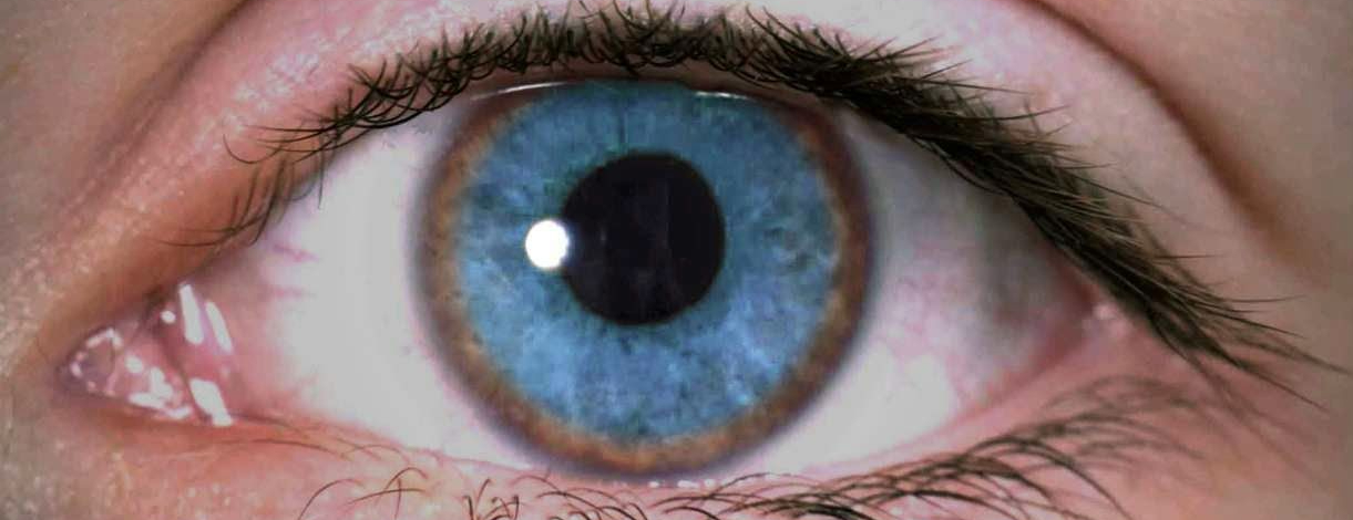
Kayser-Fleischer rings are dark circles at the periphery of the cornea in patients with Wilson disease and are considered to be fairly pathognomonic, though they usually require a slit lamp to be seen. This coupled with the fact that Wilson disease is rare means that most of us haven’t seen them.
If not Wilson, who?
Kayser-Fleischer rings were first described in 1902 when a German physician named Bernhard Kayser evaluated a patient with neurological symptoms. The patient had Wilson disease, but the disease hadn’t been described (or named) yet. Kayser thought the patient had multiple sclerosis but noted dark corneal rings in the patient’s eyes.
One year later, in 1903, a German ophthalmologist named Bruno Fleischer also described corneal rings in patients with neurological symptoms, and he took the association a step further by noting cirrhosis at autopsy.
It wasn’t until 1912 that British neurologist Samuel Kinnier Wilson described the disorder that bears his name. Wilson named it “progressive lenticular degeneration” and linked the neurological symptoms and cirrhosis, while also noting its inherited nature. Interestingly, Wilson didn’t describe the corneal rings despite the fact that his original description encompassed 212 pages in the March, 1912 issue of the journal Brain.
Anatomy refresher
To fully understand why copper deposits in the cornea, a review of corneal anatomy is required. There actually are 5 layers to the cornea.
- Epithelium on the outside
- Bowman’s layer
- Stroma
- Descemet’s membrane
- Endothelium on the inside

Descemet’s membrane is on the inner portion of the cornea, separated from the aqueous humor in the anterior chamber only by the corneal endothelium. Pathology studies of the corneas of patients with Kayser-Fleischer rings revealed that Descemet’s membrane is where the rings localize, with copper granules deposited at the periphery.
The fact that copper deposits anywhere in the cornea might come as a surprise given that it is a completely avascular structure. One clue is that pathological studies of the eyes of patients with Wilson Disease found increased copper levels in the aqueous humor. The aqueous humor in the anterior chamber of the eye is only separated from Descemet’s membrane by a thin endothelial layer. So the copper in the aqueous humor has easy access – it just has to diffuse across the endothelium and you have copper depositing in Descemet’s membrane.
But why a ring?
One might reasonably wonder why the entire cornea isn’t affected by copper deposits or why the deposits aren’t concentrated at the lower portion given the effects of gravity.
Flow dynamics of aqueous humor provide the answer. Aqueous humor flows from the posterior chamber to the anterior chamber of the eye and then drains into Schlemm’s canal at the edges of the chamber. You might think that it just flows in like a river or creek but it actually has more of a swirling configuration, which angles the flow preferentially toward the periphery.

This swirling flow leads to more contact for the copper-containing aqueous humor at the edges of the cornea and preferential deposition at the periphery. This leads to that class circular shape associated with Kayser-Fleischer rings. Arcus senilis, which are rings of cholesterol in the cornea, may have a similar mechanism.
In support of this explanation is an incredible case report from 1986. The patient had unilateral ocular trauma to the right eye when he was younger and made significantly less aqueous humor in that eye. He then developed Wilson disease but only manifested a Kayser-Fleischer ring in the left unaffected eye. This supports the role aqueous humor plays in this clinical finding!
Clinical implications
Kayser-Fleischer rings correlate well with the presence of neuropsychiatric symptoms, which are some of the dreaded complications of Wilson disease. In fact, >90% of those with neuropsychiatric symptoms have Kayser-Fleischer rings while only 50% of those who don’t have them.
Also, this findings resolves with chelation therapy as serum copper levels fall.
Take Home Points
- Kayser-Fleischer rings result from deposition of copper in Descemet’s membrane of the cornea in patients with Wilson disease
- They were first observed by Kayser and Fleischer about 10 years before Wilson formally described the disease that bears his name
- The peripheral, ringed shape of Kayser-Fleischer rings result from swirling flow of aqueous humor as it enters in the anterior chamber of the eye, which deposits copper at the edges of the cornea
- Kayser-Fleischer rings are associated with the presence of neuropsychiatric symptoms in Wilson disease and resolve as serum copper levels decrease with chelation therapy
Learning objectives
- Understand that Kayser-Fleischer rings result from deposition of copper in Descemet’s membrane of the cornea in patients with Wilson Disease
- Learn that the peripheral, ringed shape of Kayser-Fleischer rings result from swirling flow of aqueous humor as it enters in the anterior chamber of the eye, which deposits copper at the edges of the cornea
- Become aware that Kayser-Fleischer rings are associated with the presence of neuropsychiatric symptoms in Wilson disease and resolve as serum copper levels decrease with chelation therapy
CME/MOC
Click here to obtain AMA PRA Category 1 Credits™ (1.00 hours), Non-Physician Attendance (1.00 hours), or ABIM MOC Part 2 (1.00 hours).
Listen to the episode
If you want to read more, here’s a link to Avi’s tweetorial on the topic.
Credits & Citation
◾️Episode written by Avi Cooper
◾️Audio edited by Clair Morgan of nodderly.com
◾️Show notes by Tony Breu & Avi Cooper
Cooper AZ, Abrams HR, Breu AC. Why do Kayser-Fleischer rings form in the cornea in Wilson disease? The Curious Clinicians Podcast. January 6, 2021. https://curiousclinicians.com/2021/01/05/episode-16-why-do-kayser-fleischer-rings-form-in-the-cornea-in-wilson-disease/
Image credit: https://www.pinterest.dk/pin/687150855619945995/
