
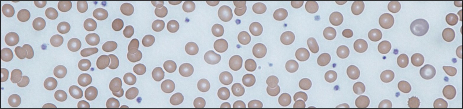
Why does iron deficiency cause thrombocytosis?
The differential for thrombocytopenia, or low platelets, is a classic. Are all the cell lines down? Usually bone marrow suppression due to anything from leukemia to Lyme disease. Is there new, severe thrombocytopenia and anemia? Make sure to rule out a microangiopathic hemolytic anemia (MAHA). Is there portal hypertension with splenomegaly? It’s probably sequestration. When platelets are low, there’s usually a reason we can find. How about when platelets are high? To be honest, most of us see thrombocytosis on a complete blood count and mutter something about it being “reactive” while hoping the number is normal on the next. Which, to be fair, is a reasonable answer, as platelets are an acute-phase reactant. But next time you see a platelet count of 600,000 on a CBC, add this to your differential: Iron deficiency.
Now what does iron deficiency have to do with platelets? And if anything, why do platelet counts get HIGHER when iron is low and hemoglobin is dropping with it? To answer that, we first need to learn more about an unsung bloodline hero: The megakaryocyte. This platelet progenitor primarily lives in the bone marrow, where it makes up only 0.5% of all cells despite being 10 to 15 times larger than the average red blood cell. Of note, it’s been known since 1893 that megakaryocytes also inhabit the lungs, but it was only in 2017 that a mouse study showed them to be highly active and likely a major contributor to total-body platelet count. Each megakaryocyte produces a few thousand platelets a day, totaling over 100 billion new platelets daily. Why so big? Unlike most precursors, which continually divide and mature, megakaryocytes actually produce platelets by breaking off parts of the cell, like flinging drops of water from an ever-running fountain.
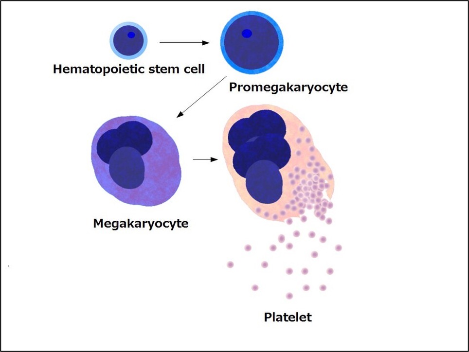
As the megakaryocyte breaks off platelets, it must constantly replicate its DNA, but not divide. It accomplishes this through endomitosis, a modified cell cycle where it begins mitosis, but doesn’t complete normal cell division by completing cytokinesis. This results in impressive polyploidy, or DNA copying. Each copy is referred to as “N,” such that a cell (like most human cells) which copies its DNA twice is “2N.” Megakaryocytes (literally “big nucleus cell” in Greek) are usually 16N, although some reach 32N or 64N. Platelets themselves don’t have nuclei or DNA (which is why they aren’t, strictly speaking, “cells”). However, each platelet needs mitochondria, alpha granules, and dense granules. These granules store polypeptides and proteins, including clotting factors, von Willebrand factor, growth factors like VEGF, and cytokines. The megakaryocyte needs all that extra DNA to continuously produce the machinery that thousands of platelets require but cannot make themselves.
Some of you may remember that platelets and red blood cells share a myeloid progenitor. For years the teaching has been that as the iron-deficient body produces more of those progenitors to help produce RBCs, some of them become megakaryocytes by accident. We now know that this is no accident. Evolutionarily, the body senses iron deficiency as a sign of bleeding. To support hemostasis, it diverts megakaryocytic-erythroid progenitors (MEPs) away from erythroid production and towards megakaryocyte production. Furthermore, even if total megakaryocytes are not increased, iron deficiency increases the polyploidy of the new megakaryocytes, making them capable of producing more platelets. The multiple mechanisms for prioritizing platelet production are the reason by thrombocytosis is seen in up to a third of iron-deficient patients, and is even associated with increased risk of thrombosis.
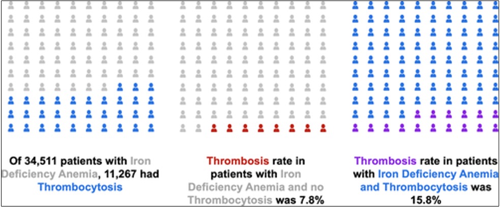
In iron deficiency, it seems like the body is acting on a “plug the drain before refilling the bath” principle. But why not do both, especially since platelets aren’t full of iron like red blood cells? It’s true that 60–70% of total body iron is incorporated into hemoglobin, making red blood cell production by far the largest consumer of this mineral. However, many enzymes in the body, especially mitochondrial ones, require iron to function. If the body responded to iron deficiency by prioritizing RBC production, total-body stores would be rapidly depleted, leaving other cells, including platelets, functioning less well. A temporary down-shift is fine, because as we learned in episode 82, we need far less hemoglobin than you’d think to maintain normal oxygen delivery.
Take Home Points
- Iron deficiency is a known cause of thrombocytosis.
- Iron deficiency leads to a megakaryocytic-erythroid progenitor lineage commitment shift towards megakaryocytes. One of the key changes is an increase in megakaryocyte ploidy.
- Platelets are produced in the lung too!
Sponsor
This episode was sponsored by FIGS. FIGS is offering 15% off your first purchase. Just go to wearfigs.com and use the code FIGSRX at checkout
Listen to the episode!
https://directory.libsyn.com/episode/index/show/curiousclinicians/id/42210725
CME/MOC
Click here to obtain AMA PRA Category 1 Credits™ (0.5 hours), Non-Physician Attendance (0.5 hours), or ABIM MOC Part 2 (0.5 hours).
As of January 1, 2024, VCU Health Continuing Education will charge a CME credit claim fee of $10.00 for new episodes. This credit claim fee will help to cover the costs of operational services, electronic reporting (if applicable), and real-time customer service support. Episodes prior to January 1, 2024, will remain free. Due to system constraints, VCU Health Continuing Education cannot offer subscription services at this time but hopes to do so in the future.
Credits & Suggested Citation
◾️Episode written by Tony Breu
◾️Show notes written by Tony Breu and Giancarlo Buonomo
◾️Audio edited by Clair Morgan of nodderly.com
Breu AC, Abrams HR, Cooper AZ, Buonomo G, Manna, M. Ironing Out More Platelets. The Curious Clinicians Podcast. July 25th, 2026.
Image Credit : Wikimedia

